Back
Analysis of the surface of a reference sample made of Go SiFe steel
The ESIEE Amiens, the UPJV-LTI (Laboratory of Innovative Technologies) and the IRT-M2P carry out metallographical, optical and magnetic surface analysis on FeSi GO reference samples.
The IRT-M2P (Institute of Technological Research – Materials, Metallurgy, Processes) uses a revelation technique of the grains by chemical etching, and then by optical microscopic observation. Then, ESIEE uses an atomic force microscope, equipped with a magnetic sensor to conduct Magnetic Force Microscopy (MFM) imaging experiments. Finally, ESIEE, outsources magneto-optical observations of magnetic domains to the EVICO MAGNETICS company. Two techniques are used:
- MOIF: Magneto-Optic Imaging Film technique
- MOKE: Magneto-optical Microscopy by Kerr Effect in reflection.
Both techniques use the physical phenomenon of the light polarization rotation during its reflection on some magnetic media, since the direction and intensity of the rotation depend on the orientation and intensity of the magnetic polarization.
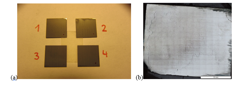
Figure 1: (a) samples of 10*10 mm2 for MOIF and MOKE Imaging (b) Revelation of grain joints in metal.
Grain oriented electrical steels are known for having huge sized-grains, that can reach more than 1 cm (see Figure 1). This peculiarity also results in relatively big domains, as we will see in the following observations.
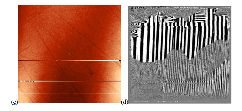
Figure 2: (c) phase magnetic contrast observed by MFM with a hard-magnetic sensor (scan size < 120*120 mm2) (d) magnetic domains observed by MOKE (scan size < 10*10 mm2).
The study of the surface magnetic structure of the grain-oriented silicon steel samples provided by SEPSA provides key information on the distribution in magnetic domains, as well as their average size. The atomic force microscope in MFM mode (Magnetic Force Microscopy) used by ESIEE and UPJV only enables to investigate areas with a surface inferior to 120*120 μm2 (see Figure 2 (c)). Considering the significant size of magnetic domains, this observation area only allows the non-exhaustive observation of three domains (see the light and dark areas on Figure 2 (c)). However, it is possible to deduce an approximative average size of magnetic domains in zero field. This distribution and distinctive size seem coherent with the observations made through the MOIF and MOKE techniques (see Figure 2 (d), made with the help of the EVICOMAGNETICS company and Mr. Rudolf Schaefer), where the investigated area is much more important (60 times the scan size of the MFM). Between 50 and 100 domains appear on this figure, which enables an estimation of the magnetic domains’ minimal and maximal sizes, which is in the range of the domain size obtained by MFM.